
Daily activities become a challenge when knee cartilage loses its smooth, shock-absorbing surface. Emerging non-surgical strategies—ranging from targeted exercise to regenerative biologics—now offer realistic hope for restoring comfort and function without the risks or downtime of an operating room. Below is a deep dive into the science, practical steps, and future innovations that can help you move freely again.
1. What Happens When Cartilage Breaks Down?
Articular cartilage is an ultra-slick layer of hyaline tissue that coats the ends of your femur and tibia, enabling near-frictionless motion. Because it lacks its own blood supply, wear-and-tear or injury heals poorly, often progressing to osteoarthritis. MRI signs include surface fibrillation, focal defects, and joint-space narrowing. Over time, biomechanical overload triggers inflammation, swelling, and pain that disrupt gait and degrade surrounding ligaments and menisci. PMC
2. Why Avoid Surgery if Possible?
- Lower complication risk: No anesthesia, infection, or thrombosis concerns.
- Faster return to activity: Most evidence-based protocols let you resume light exercise in days, not months.
- Cost efficiency: Out-of-pocket expenses for physical therapy or biologic injections are often lower than arthroscopic repair or knee replacement.
- Preservation of native tissue: Non-invasive approaches focus on stimulating intrinsic healing rather than cutting or removing structures.
Surgery remains vital for large, full-thickness lesions or severe malalignment, yet many patients qualify for conservative care when cartilage loss is mild-to-moderate.
3. Build a Resilient Musculoskeletal Foundation
3.1 Joint-Friendly Movement
- Closed-chain kinetics: Mini-squats, wall sits, and stationary cycling generate compressive “pump” forces that ferry nutrients into avascular cartilage.
- Aquatic therapy: Buoyancy unloads joints while resistance from water strengthens quadriceps and gluteal stabilizers.
- Neuromuscular training: Balance boards and single-leg stands refine proprioception, reducing aberrant shear that erodes cartilage.
Consistency is critical; even 15-minute sessions, five days a week, can bolster cartilage glycosaminoglycan content in as little as 12 weeks, according to longitudinal MRI studies. PMC
3.2 Flexibility & Fascia Release
Tight iliotibial bands or calf muscles increase patellofemoral pressure. Use foam rolling, dynamic stretches, and yoga-style poses (e.g., low lunge) to lengthen tissue and distribute load evenly across the knee.
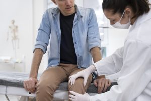
4. Regenerative Injection Therapies
Note: Most biologic interventions remain under active investigation. Efficacy, dosing, and long-term safety are not yet standardized; discuss individualized suitability with a qualified clinician.
4.1 Platelet-Rich Plasma (PRP)
PRP concentrates a patient’s own growth factors (TGF-β, PDGF, VEGF) to spark chondrocyte proliferation and dampen inflammation. Meta-analyses show modest pain reduction and functional gains for up to 12 months compared with saline or corticosteroid injections. PMC
4.2 Mesenchymal Stem Cell (MSC) Injections
Bone-marrow- or adipose-derived MSCs can differentiate into chondrocyte-like cells and secrete trophic factors that remodel extracellular matrix. Early-phase trials reveal MRI evidence of defect fill and improved KOOS scores, though heterogeneity in cell dose and culture methods complicates comparisons. DVC Stem
4.3 Exosome-Enhanced Formulations
Cell-free nanopackets of RNA and proteins harvested from PRP or MSC cultures may bypass regulatory hurdles associated with live cells. Preclinical rabbit models show thicker cartilage and higher type II collagen content when exosomes are combined with calcium-phosphate scaffolds. Human data remain scarce—flag for ongoing review as trials mature. BioMed Central
4.4 Injectable Hydrogels and Bioactive Scaffolds
Next-generation hydrogels, sometimes loaded with chondrogenic peptides or gene vectors, can be injected arthroscopically or via needle to create a 3-D matrix that “molds” to defect topology. Northwestern University’s bioactive gel regenerated high-quality cartilage in large-animal knees eight months post-injection, a milestone that may translate into human trials soon.
5. Lifestyle & Nutritional Levers
5.1 Weight Management
Every additional kilogram transmits up to four kilograms of force across the knee when walking. Even a 5 % body-weight reduction lessens symptomatic severity and delays radiographic progression.
5.2 Anti-Inflammatory Eating Pattern
Prioritize omega-3-rich fish, colorful polyphenol-dense produce, and spices such as turmeric. Limit ultra-processed foods and added sugars that fuel systemic inflammation.
5.3 Joint-Support Supplements (Evidence quality: mixed; verify tolerability)
| Compound | Proposed Action | Typical Course |
|---|---|---|
| Glucosamine Sulfate | Substrate for glycosaminoglycan synthesis | 1500 mg daily, ≥3 months |
| Chondroitin | Inhibits degradative enzymes | 800–1200 mg daily |
| Undenatured Collagen (UC-II) | Oral tolerance to reduce immune attacks on cartilage | 40 mg daily |
6. External Supports & Device-Based Modalities
- Unloader braces: Shift tibiofemoral contact away from damaged compartment; clinical trials show 10–15 % pain relief.
- Shock-absorbing insoles: Dampen ground-reaction forces; best for high-impact occupations or sports.
- Blood-flow-restriction (BFR) bands: Low-load resistance training under partial occlusion induces hypertrophy comparable to heavy lifting, protecting sensitive joints.
- Low-level laser and pulsed ultrasound: May enhance mitochondrial function and microcirculation; guidelines remain inconsistent on dose and frequency.
- Extracorporeal shockwave therapy (ESWT): High-energy pulses stimulate subchondral bone remodeling; preliminary data indicate gait-speed gains but more randomized trials are needed.
7. Cutting-Edge Horizons (2024 – 2025)
- Nasal-cartilage cell grafts – Harvesting septal chondrocytes, expanding them in vitro, and percutaneously injecting engineered tissue is being studied as a less invasive alternative to osteochondral graft surgery. Early cohorts report >90 % mobility improvement at one year. Latest news & breaking headlines
- Gene-activated hydrogels – Carrier matrices releasing BMP-7 or SOX-9 plasmids encourage in-situ chondrogenesis; animal work shows robust collagen type II deposition for up to 12 months. PMC
- 3-D bioprinted micro-scaffolds – Portable printers can produce patient-specific, injectable constructs seeded with MSCs at point of care, potentially streamlining regulatory pathways.
- Exosome-loaded adhesives – Hybrid hydrogels doped with vesicles adhere to wet cartilage, resisting shear better than standard fibrin glues and sustaining cytokine release over weeks. ScienceDirect
Many of these innovations are confined to animal or early human feasibility studies. Rigorous, adequately powered clinical trials must confirm durability, cost-effectiveness, and scalability before routine use.
8. Who Benefits Most From Non-Surgical Cartilage Repair?
| Candidate Profile | Likely Response |
|---|---|
| Adults under 60 with localized grade II–III defects (<2 cm²) | High – native repair capacity still reasonable |
| Early-stage osteoarthritis with BMI < 35 | Moderate – biologics + weight loss can slow degeneration |
| Advanced tricompartment OA or severe limb malalignment | Low – surgical realignment or arthroplasty often required |
A thorough orthopedic assessment—including weight-bearing X-rays, high-resolution MRI, and movement analysis—helps tailor a multimodal plan.
9. Practical Roadmap to Renewed Mobility
- Baseline evaluation: Imaging, biomechanical screening, and blood tests for metabolic factors.
- Three-month conservative block: Integrated physical therapy, nutrition overhaul, and bracing.
- Regenerative add-ons: If pain or function plateaus, layer PRP or MSC-based injections.
- Reassess with MRI at six months: Track defect fill and edema resolution.
- Advance to higher-impact sport or work duties once strength and proprioception meet protocol targets.
- Monitor annually: Adjust exercise volume, nutrition, and supportive devices as needed.
10. Key Takeaways
Non-surgical knee cartilage repair is no longer limited to “cope and wait.” Evidence-backed movement programs, strategic weight control, and increasingly sophisticated biologic injectables can synergize to reduce pain, rebuild tissue architecture, and delay—or potentially prevent—major surgery. Staying informed about emerging hydrogels, exosome technologies, and cell-based innovations will maximize your odds of stepping into a future with healthier, happier knees.